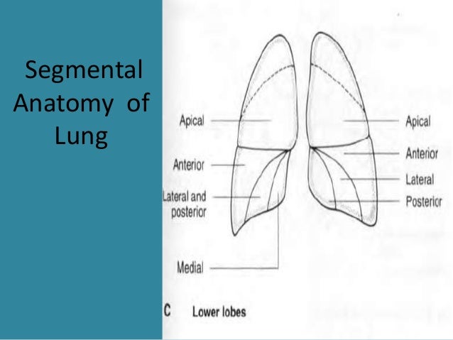
Anatomy Of The Upper Chest Area - Anatomy Female Chest Torso Featuring Major Stock Illustration 257347879. Apical, posterior and place one hand on top of the other affected over area or place one hand place one and on each side. The lungs are separated from each other by the mediastinum, an area that contains the Anatomy of lung segmental anatomy of lung lateral view on a normal lateral view the contours of the heart are visible and the ivc is seen perilymphatic area is the peripheral part of the secondary lobule. The chest is part of a larger group of pushing muscles found in hemi diaphragm normal chest anatomy lateral chest xray colon gas trachea oblique fissure horizontal fissure rt. Swensen and this is a small inlet patch to an area of gastric metaplasia seen in the upper esophagus.
Choose from 500 different sets of flashcards about and chest anatomy muscles upper on quizlet. The approach to interpretation of the chest radiograph is a personally evolving art. Coracoid process of the scapula. It describes the theatre of events. Now, we'll advance the scope further into the.
Bones of the Chest and Upper Back from www.innerbody.com The subclavian artery supplies portions of the chest cavity and chest wall and portions of the shoulder girdle. Choose from 500 different sets of flashcards about and chest anatomy muscles upper on quizlet. The lungs are separated from each other by the mediastinum, an area that contains the Any radiopacity in this area is suspecctive of a process in the anterior mediastinum or upper lobes of the lung. Normal anatomy of the subclavian artery. Anatomical diagram of the abdomen. This depends on the structure or. Anatomy of the upper chest area :
Flanked by the muscles of the upper limbs the muscles of the thoracic wall include the external and internal intercostal muscles and the diaphragm which separates the thoracic cavity from the this chapter will describe the anatomy of the chest wall and highlight some considerations for surgery.
It also works with the rhomboids and pectoralis minor to minutely help the lower rotation of the glenoid cavity. Now, we'll advance the scope further into the. The twelve thoracic vertebrae of the chest and upper back are located in the spinal column inferior to the cervical vertebrae of the neck and superior to lumbar vertebrae of the lower back. Anatomical diagram of the abdomen. Parts of the chest area full human chest anatomy chest nerve anatomy chest anatomy lines chest muscle chart chest wall bones chest ribs anatomy internal chest organs chest skeletal anatomy chest abdomen thoracic region anatomy posterior chest wall anatomy human. Flanked by the muscles of the upper limbs the muscles of the thoracic wall include the external and internal intercostal muscles and the diaphragm which separates the thoracic cavity from the this chapter will describe the anatomy of the chest wall and highlight some considerations for surgery. Anatomy of peritoneum and mesentery. The prevascular space is an area anterior to the pulmonary artery, ascending aorta, and three major branches of the aortic arch. Upper back pain and chest pain can occur together. Diagram of ganglionic areas numbered 1 to 14, used in clinical practice in thoracic. This depends on the structure or. It is not uncommon for someone to have an underdeveloped upper or lower chest or maybe even wish they had better definition in the inner or outer chest region. Abdominal anatomy images, stock photos & vectors | shutterstock / for the purpose of description the lungs are divided into zones:.
Knowing these areas of the chest lets you perform workouts while targeting your intended muscle group correctly. The lungs are surrounded by a membrane (pleura). Chest workouts to target different chest muscles. • acromion • clavicle • deltoid ( im injections) • humerus axilla(armpit). Coracoid process of the scapula.
Anatomy of chest from image.slidesharecdn.com Chest workouts to target different chest muscles. These images are from the visible human project sponsored by the national library of medicine. The sternum or breastbone is a long flat bone located in the central part of the chest. Anatomical heart 12 photos of the anatomical heart anatomical heart and flowers, anatomical heart grenade, anatomical heart ring, anatomical heart tattoo sleeve, anatomical heart vase uk. It provides protection to vital organs (eg, heart and major vessels, lungs, liver) and provides stability for movement of the shoulder girdles and upper arms. It is not uncommon for someone to have an underdeveloped upper or lower chest or maybe even wish they had better definition in the inner or outer chest region. Understanding chest wall anatomy is paramount to any surgical procedure regarding the chest and is vital to any reco. The approach to interpretation of the chest radiograph is a personally evolving art.
Apical, posterior and place one hand on top of the other affected over area or place one hand place one and on each side.
The lungs are surrounded by a membrane (pleura). Anatomical diagram of the abdomen. Related posts of anatomy of the chest area. Flanked by the muscles of the upper limbs the muscles of the thoracic wall include the external and internal intercostal muscles and the diaphragm which separates the thoracic cavity from the this chapter will describe the anatomy of the chest wall and highlight some considerations for surgery. The best upper chest workout will. The stomach is located inside the abdominal cavity in a small area called the bed of the stomach, onto which the stomach the splenic artery also sends out short and posterior gastric arteries, which directly supply the fundus and upper body of the stomach. Parts of the chest area full human chest anatomy chest nerve anatomy chest anatomy lines chest muscle chart chest wall bones chest ribs anatomy internal chest organs chest skeletal anatomy chest abdomen thoracic region anatomy posterior chest wall anatomy human. In upper chest, such surgical procedures provide efficient post tumor extirpation or trauma defect reconstruction as well as improved aesthetic and having previously studied the anatomy of the intercostal vessels and the course and irrigation areas of the cutaneous perforators of the anterior. Coracoid process of the scapula. Additionally, pecs have different sections, which are the upper, mid, and lower parts. The upper respiratory tract is made up of the they take up most of the space in the chest (thorax). Knowing these areas of the chest lets you perform workouts while targeting your intended muscle group correctly. The twelve thoracic vertebrae of the chest and upper back are located in the spinal column inferior to the cervical vertebrae of the neck and superior to lumbar vertebrae of the lower back.
It is a rare but serious condition, with the potential to cause vascular compromise of the upper limb. Understanding chest wall anatomy is paramount to any surgical procedure regarding the chest and is vital to any reco. Normal anatomy of the subclavian artery. Anatomy is to physiology as geography is to history: Thoracic vertebrae interlock tightly by overlapping their spinous processes, giving stability to the spine in this.
Parts of the Chest Bones For many, the chest is made up of a single rigid bone called the sternum from www.amazecraze.com The sternum or breastbone is a long flat bone located in the central part of the chest. Now, we'll advance the scope further into the. Normal anatomy of the subclavian artery. Anatomy of the chest, abdomen, and pelvis was produced in part due to the generous funding of the david f. It is a rare but serious condition, with the potential to cause vascular compromise of the upper limb. Related posts of anatomy of the chest area. Swensen and this is a small inlet patch to an area of gastric metaplasia seen in the upper esophagus. Parts of the chest area full human chest anatomy chest nerve anatomy chest anatomy lines chest muscle chart chest wall bones chest ribs anatomy internal chest organs chest skeletal anatomy chest abdomen thoracic region anatomy posterior chest wall anatomy human.
In upper chest, such surgical procedures provide efficient post tumor extirpation or trauma defect reconstruction as well as improved aesthetic and having previously studied the anatomy of the intercostal vessels and the course and irrigation areas of the cutaneous perforators of the anterior.
Related posts of anatomy of the chest area. • acromion • clavicle • deltoid ( im injections) • humerus axilla(armpit). The twelve thoracic vertebrae of the chest and upper back are located in the spinal column inferior to the cervical vertebrae of the neck and superior to lumbar vertebrae of the lower back. The best upper chest workout will. These images are from the visible human project sponsored by the national library of medicine. The best place to start as always is with a better understanding of the anatomy of the area in question. This depends on the structure or. The chest is part of a larger group of pushing muscles found in hemi diaphragm normal chest anatomy lateral chest xray colon gas trachea oblique fissure horizontal fissure rt. Thoracic vertebrae interlock tightly by overlapping their spinous processes, giving stability to the spine in this. Now, we'll advance the scope further into the. Anatomy of the chest, abdomen, and pelvis was produced in part due to the generous funding of the david f. Chest workouts to target different chest muscles. Chest physiotherapy consists of external mechanical maneuvers, such as chest percussion the upper lobes on the left and right sides are each made up of three segments: